| |
FemaleHealthMadeSimple | |
|
CLICK A TOPIC TO VIEW IT
HOME
A NEW LIFE BEGINS
VERY EARLY PREGNANCY
THE PILL AND OTHER FORMS
ABNORMAL MENSTRUATION
ENDOMETRIOSIS
FEMALE GROWTHS AND CANCER
GYNECOLOGICAL OPERATIONS
SONARS
PUBERTY
MENOPAUSE
PREGNANCY AND CHILD BIRTH
GLOSSARY
|
Do You Understand Your Body?What is a Sonar? or a Ultrasound? Why do doctors do sonar examinations?
SONAR EXAMINATIONS AND THEIR VALUE
Sonar . The word or thought brings forward visions of images of unborn babies .
We will discuss it's uses in both gynecology and pregnancy. The term SONAR is derived from Sound Navigation and Ranging. High frequency sound waves are transmitted into the body . Different tissues reflects the sound waves differently and the reflected sound waves (echoes) are used to create the sonar image. The machines used in medical applications use very high frequency sound waves and are therefore also referred to as ultrasound machines. The part of the sonar machine that touches the body is called a probe. It is a very complicated piece of equipment. It contains crystals that are able to transform electrical energy into high frequency sound waves. The same crystals are also able to to the opposite and change sound waves into electrical energy. The probe sends a short burst of high frequency sound waves and than listens for the return echoes . The time that elapses between transmission and reception indicates the distance from the sonar probe to the object that is examined. The crystals are technically manipulated in such a way that a two dimensional picture is generated on a screen (monitor). Just high frequency sound waves are generated and no rays.
Even if it is just sound waves , is it save? High frequency sound waves do shake molecules around
and can generate heat in tissues. The sound waves are transmitted in short bursts of milliseconds
duration and for most of the period that the examination lasts, the probe is listening and
interpreting the returning echoes. The returning echoes are much weaker and it is very
doubtful that they will have any harmful effect on tissues. We can never guarantee
100 % safety but it is our view that sonar examinations are very save.
A major advance in the gynecological application of sonar examinations was the advent of the transvaginal probe. The clarity is better and there is no need for a full bladder. For the pelvic organs to be visualized with a transabdominal abdominal probe ( a probe touching the tummy) the bladder must be very full, causing extreme discomfort. The vaginal probe is a thin tube that easily fits in the vagina. It is covered by a condom before insertion into the vagina. The tip of the probe is very near the female organs and a clear sonar image is obtained. The tip of the probe is also visible helping the examiner to accurately orientate himself or herself to the area being examined. The thickness of the endometrium ( the lining covering the cavity in the womb) is easily measured and it's thickness is giving valuable information. It's appearance on a transvaginal sonar differs at different stages of the menstrual cycle. The sonar photos shown on this page are real but the drawings are very schematic and are just intended to supplement the sonar photos. Click here to refer to an explanation of the changes occurring in the endometrium during the monthly cycle. The lining should be thin during the first few days following the menstruation. (fig 1 and fig 2)
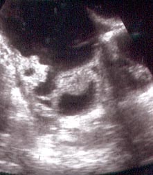
A thin endometrium is only normal during the first few days after a mestruation stopped or after the menopause. At any other time during the cycle a thin endometrium is an indication of a lack of estrogen. The lining has a typical appearance during the middle part of the cycle.(7 to 14 days after the mestruation). (fig 3 and fig 4)
This type of appearance is only normal during the middle of cycle before ovulation occurs. (the second week of the cycle), At any other time it is an indication of a lack of progesterone and highly suspicious of the absence of ovulation. During the latter part of the cycle the endometrium has another typical appearance illustrated in fig 5 and 6.
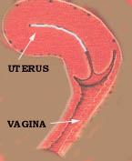
This appearance is an indication that the cycle is completely normal and that ovulation occurred. It represents a normal cycle at about 21 days after the previous menstruation. < P> Refer to normal menstruation to refresh your memory about the different stages of the menstrual cycle. A transvaginal sonar is also of great value value in examining the ovaries. The following photos illustrate the appearance of the ovaries using a transvaginal ultrasound probe under different conditions.
Photo A
Photo B
< pco slater cyst>
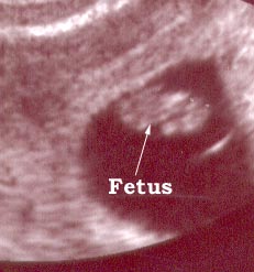
Photo C This is a ovary with multiple irregular cysts and turned out to a cystic cancer of the ovary. This photo is included to stress the importance of regular checks. Regular annual examinations and regular ultrasound examinations done through the vagina might help to diagnose ovarian cancer early. The transvaginal sonar also give an excellent view of the cervix ( mouth of the womb) helping doctors to excess conditions effecting the cervix. We tried to explain the increasing importance of a transvaginal ultrasound (sonar) examinations in assisting doctors to access female (gynecological) diseases. We will now discuss the use of ultrasound in pregnancy. The first concern is the safety to the fetus. Until now no dangerous side effects or abnormalities caused by sonar examination has been discovered. The role of sonar examinations during pregnancy is to detect the duration of pregnancy (dating the pregnancy), to detect fetal abnormalities and to monitor fetal development and growth. It is important to understand that the sonar is just a machine and can't determine the duration of pregnancy. What actually happens is that different fetal parts are measured and that according the size of the specific part a computer program calculates the duration of the pregnancy. The programs are developed by doing thousands of ultrasound examinations at different stages of pregnancy. The accuracy of sonar dating of a pregnancy depends on different factors. Is the program appropriate to the community where it is used. If the program was developed in a country where the average fetal size is small and used in community where the average fetal size is large ( or vice versa) unreliable results will be obtained. A fetus in the population with the large average size will appear to bigger and older than what it actually is. The opposite will happen in the population with the smaller than average size fetus. The size of a fetus also influences the accuracy of a ultrasound's estimation of the duration of pregnancy. A larger than average fetus will cause the program to abnormally increase the duration and a small fetus will cause the opposite effect. Here are a few sonar photos to illustrate the appearance of the fetus at different stages of a pregnancy.
Photo One
Photo Two
Photo Three
Another point to remember is that the reference point used on ultrasound machines to determine the duration of pregnancy is the first day of the last menstruation and NOT THE MOMENT OF CONCEPTION.
There are two reference points used to determine the duration of pregnancy :
The fist is in use for centuries because the only known fact was that menstruation stopped during pregnancy Than a new scientific division known as embryology developed. Embryology is the study of development before birth. Embryologists originally examined animal embryos with a known date of conception. Hence the moment of conception became their reference point. Human embryologists also use the moment of conception as their reference point, Obstetricians however still use the first day of the last menstruation as their reference point. This can lead to confusion because conception occurs about 2 (two) weeks after the first day of the last menstruation. The reference point use also has legal implications in fraternity disputes. If a sonar examination indicates a twelve week pregnancy , the duration is only 10 weeks and conceptions occurred about 10 weeks ( and not twelve weeks) previously. |
Go to the top of the page.
LAST UPDATE : 27 December 2002
Copyright FemaleHealthMadeSimple 2001
Did you ever wonder why people buy stuff over the internet? Do they really buy stuff?
Visit an internet mall and decide for yourself.
Click here and go to a cyber mall.
Music,books and magazines. Just click here.


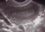
 A normal ovary. A few small follicles are visible ( it is completely normal)